Early childhood depression alters brain development
Findings may help explain why those who are depressed have difficulty regulating their moods and emotions

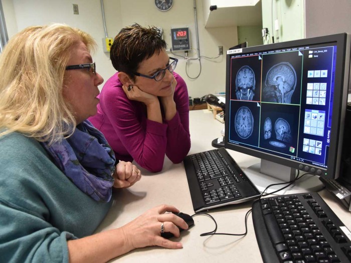
Deanna M. Barch, PhD, (left) and Joan L. Luby, MD, examine brain images for differences in brain tissue between children depressed as preschoolers and those who were not. They have linked preschool depression with less gray matter and the thinning of that tissue in the brain’s cortex. <em>Photo: Robert Boston</em>

The brains of children who suffer clinical depression as preschoolers develop abnormally, compared with the brains of preschoolers unaffected by the disorder, according to new research at Washington University School of Medicine in St. Louis.
Their gray matter — tissue that connects brain cells and carries signals between those cells and is involved in seeing, hearing, memory, decision-making and emotion — is lower in volume and thinner in the cortex, a part of the brain important in the processing of emotions.
The new study is published Dec. 16 in JAMA Psychiatry.
“What is noteworthy about these findings is that we are able to see how a life experience — such as an episode of depression — can change the brain’s anatomy,” said first author Joan L. Luby, MD, whose research established that children as young as 3 can experience depression. “Traditionally, we have thought about the brain as an organ that develops in a predetermined way, but our research is showing that actual experience — including negative moods, exposure to poverty, and a lack of parental support and nurturing — have a material impact on brain growth and development.”
The findings may help explain why children and others who are depressed have difficulty regulating their moods and emotions. The research builds on earlier work by Luby’s group that detailed other differences in the brains of depressed children.
Luby, the Samuel and Mae S. Ludwig Professor of Child Psychiatry, and her team studied 193 children, 90 of whom had been diagnosed with depression as preschoolers. They performed clinical evaluations on the children several times as they aged. The researchers also conducted MRI scans at three points in time as each child got older. The first scans were performed when the kids were ages 6 to 8, and the final scans were taken when they were ages 12 to 15. A total of 116 children in the study received all three brain scans.
“If we had only scanned them at one age or stage, we wouldn’t know whether these effects simply were present from birth or reflected an actual change in brain development,” said co-investigator Deanna M. Barch, PhD, head of Washington University’s Department of Psychological and Brain Sciences in Arts & Sciences. “By scanning them multiple times, we were able to see that the changes reflect an actual difference in brain maturation that emerges over the course of development.”
The gray matter is made up mainly of neurons, along with axons that extend from brain cells to carry signals. The gray matter processes information, and as children get older, they develop more of it. Beginning around puberty, the amount of gray matter begins to decline as communication between neurons gets more efficient and redundant processes are eliminated.
“Gray matter development follows an inverted U-shaped curve,” Luby said. “As children develop normally, they get more and more gray matter until puberty, but then a process called pruning begins, and unnecessary cells die off. But our study showed a much steeper drop-off, possibly due to pruning, in the kids who had been depressed than in healthy children.”
Further, the steepness of the drop-off in the volume and thickness of the brain tissue correlated with the severity of depression: The more depressed a child was, the more severe the loss in volume and thickness.
The researchers determined that having depression was a key factor in gray matter development. In scans of children whose parents had suffered from depression — meaning the kids would be at higher risk — gray matter appeared normal unless the kids had suffered from depression, too.
Interestingly, the differences in gray matter volume and thickness typically were more pronounced than differences in other parts of the brain linked to emotions. Luby explained that because gray matter is involved in emotion processing, it is possible some of the structures involved in emotion, such as the brain’s amygdala, may function normally, but when the amygdala sends signals to the cortex — where gray matter is thinner — the cortex may be unable to regulate those signals properly.
Luby and Barch are planning to conduct brain scans on even younger children to learn whether depression may cause pruning in the brain’s gray matter to begin earlier than normal, changing the course of brain development as a child grows.
“A next important step will involve determining whether early intervention might shift the trajectory of brain development for these kids so that they revert to more typical and healthy development,” said Barch, also the Gregory B. Couch Professor of Psychiatry.
Luby said that is the main challenge facing those who treat kids with depression.
“The experience of early childhood depression is not only uncomfortable for the child during those early years,” she said. “It also appears to have long-lasting effects on brain development and to make that child vulnerable to future problems. If we can intervene, however, the benefits might be just as long-lasting.”


