Non-invasive imaging can pinpoint irregular heartbeat
Imaging technology developed at Washington University School of Medicine can help pinpoint electrical trouble spots in the heart

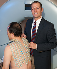
Components of an Electrocardiographic imaging (ECGI) vest that can detect electrical activity in the heart.
An irregular heartbeat, or arrhythmia, is unique in every patient and requires precise diagnosis and treatment. Electrocardiographic imaging (ECGI), an exciting advance in noninvasive cardiac imaging, can map an irregular heart rhythm in a single beat.
Invented by Yoram Rudy, PhD, the Fred Saigh Distinguished Professor of Engineering, professor of medicine, and director of the Cardiac Bioelectricity and Arrhythmia Center at Washington University, the vest-like device uses 256 electrodes combined with rapid CT image scanning to display a patient’s cardiac electrical activity in great detail. With accuracy of about six millimeters, the technology can pinpoint sites in the heart that initiate an arrhythmia.
Less invasive
Unlike invasive catheter studies, ECGI can map the arrhythmia in a single beat, and it does so without the associated risk of having to place a catheter in the heart, itself. ECGI also has the ability to image regions of the heart associated with heart attacks and the development of sudden cardiac death. Physicians also hope to use ECGI to identify high-risk patients before they have had a life-threatening arrhythmic event.

“The information provided by this technology may eventually help physicians plan individualized medical treatment,” says Phillip Cuculich, MD, one of seven Washington University electrophysiologists at Barnes-Jewish Hospital. Cuculich has worked with ECGI since 2006. “For patients, this ultimately translates into safer treatment plans with better cure rates.”


