Restoring hearing loss: Chick studies show promise
New research in chicks is identifying factors involved in regenerating the inner ear hair cells. This work could lead to new therapies for hearing loss

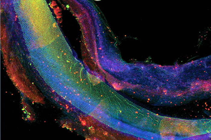
The chick cochlea contains 10,000 sensory hair cells (shown in green) which detect sound in a very similar fashion to their counterparts in the human ear. But unlike human sensory cells, the chick cells can quickly regenerate if damaged. Understanding the biologic basis of this repair process may lead to methods of inducing regeneration in the human ear.
Researchers at Washington University School of Medicine have identified the specific genes involved in regenerating, or regrowth of, cochlear hair cells in the ears of chicks — a significant step in the quest to reverse hearing loss in humans after inner ear hair cell damage.
Acoustic trauma, infections, drugs toxic to the ear and aging can take a toll on the roughly 12 thousand sensory hair cells that are present in a normal human cochlea at birth.
A dwindling stock of cochlear sensory hair cells is responsible for some degree of hearing loss for approximately a third of the US population between age 65 and 75, and affects up to half of those over 75.
Although this phenomenon is common in humans and other mammals, it is not the case with other vertebrates such as fish, birds, amphibians and reptiles. Studies of birds, for example, have shown that damaged cochlear hair cells can be replaced in as few as two to three days, with full hearing restored within a few weeks. Washington University School of Medicine researcher Mark Warchol, PhD, a professor of otolaryngology, has spent more than 20 years investigating how they do it.
“It’s likely that regeneration is probably the “default” condition of the vertebrate ear that was lost during the course of mammalian evolution,” said Warchol.
He notes that a limited degree of regeneration following injury is possible during early development of the mammalian ear. The cochlea, however, has no regenerative ability. Nevertheless, Warchol and others believe that it may be possible to induce some amount of cochlear repair through the use of drugs or genetic manipulation.
Warchol is one of a handful of researchers nationwide investigating the molecular details of cochlear development to identify specific pathways that permit and/or restrict regeneration.
Genetic involvement in regeneration
Along with colleagues in the United Kingdom, Warchol’s lab is using an advanced genetic screening technique called RNA sequencing, provided by Washington University’s Genome Institute, to identify the specific genes that are turned on (or off) in the regenerating cochlea.
With this technique, Warchol has been able to characterize the changes that follow hair cell injury by comparing gene function in regenerating and non-regenerating ears. “We compared RNA from a damaged (with antibiotic) and an undamaged chick ear at 24-hour intervals to understand what genes are turned off or on,“ said Warchol. “We now have a blueprint of the pathways involved in chick hair cell regeneration.”
Warchol has shared his findings with others in the field. He is currently using the blueprint to identify the early switches involved in the pathways by meticulously examining the functions of various genes of interest. An example is his investigation with Washington University developmental biologist David Ornitz, MD, PhD, of a fibroblast growth factor, FGFR1, identified as an essential signal that regulates the growth of cochlear sensory hair cells and supporting cells.
“This growth factor is present in the developing mammalian ear, but stops being expressed once the cochlea is mature,” said Warchol. “In chickens the gene remains turned on for the life of the animal. This may be one reason why the chicken ear is able to regenerate. We need to know more about how this occurs.”
The work by Warchol and his colleagues has moved the field toward a more complete representation of the regulatory network involved in regeneration of inner ear hair cells. This level of understanding is essential to development of therapeutic repair or regeneration of lost or damaged sensory receptors in the inner ear.


