School of Medicine scientists develop iBooks for medical classes
Educational multimedia elements mimic a microscope's view
 Robert Boston
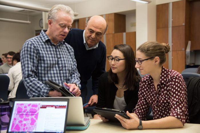
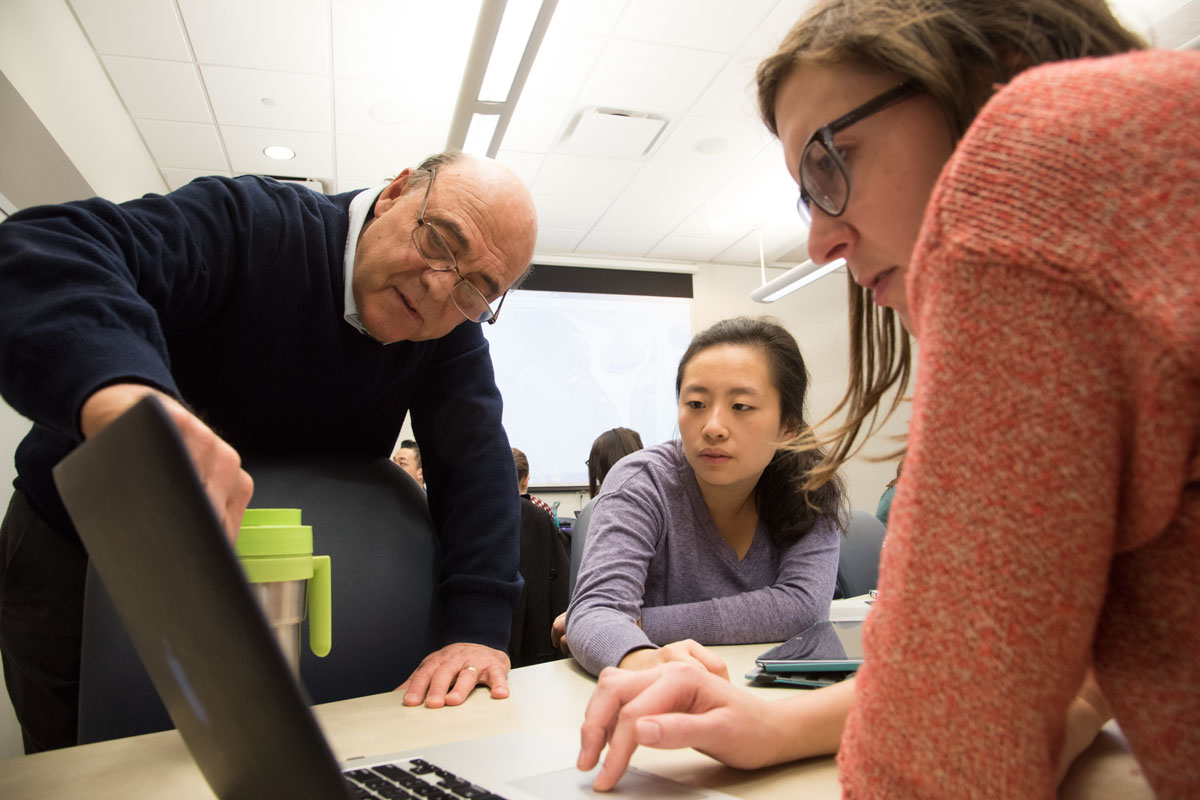
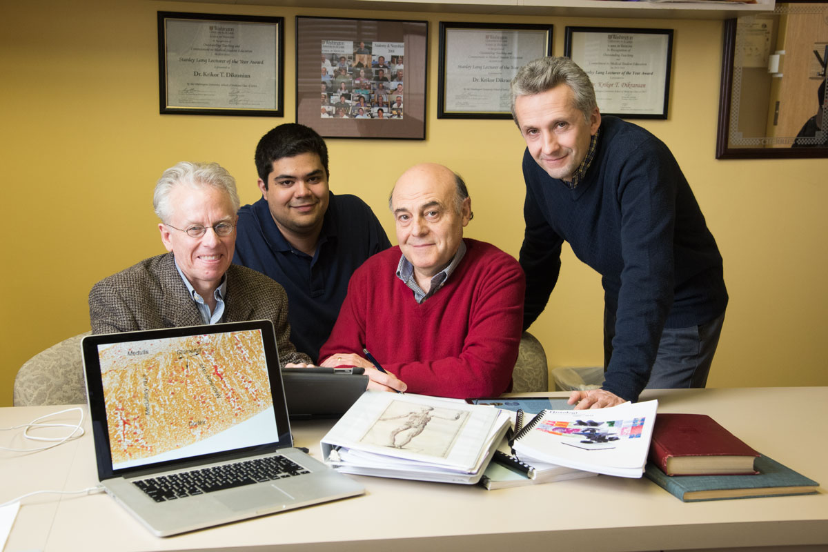
From left, Paul Bridgman, PhD, and Krikor Dikranian, MD, PhD, professors in the Department of Neuroscience at Washington University School of Medicine in St. Louis, demonstrate the histology iBook they created to medical students Caroline Min and Sarah Tepper.
Professors in the Department of Neuroscience at Washington University School of Medicine in St. Louis have developed multimedia medical books for teaching, with high-resolution slides that illustrate microscopic anatomy so precisely, it’s like looking through a microscope.
Unlike most other digital textbooks, these were created specifically as iBooks with distinctive educational features such as narrated virtual slide-viewing tutorials, integrated self-quizzes and links to online resources that save readers from having to log in to other sites. The books’ high-tech capabilities have captured the interest of education specialists at Apple, said co-author Krikor Dikranian, MD, PhD, a professor of anatomy.
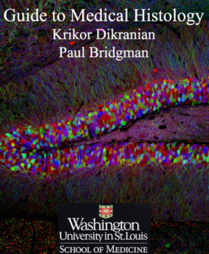
Inspired to create a stronger connection between traditional medical courses and students accustomed to all things digital, Dikranian and co-author Paul Bridgman, PhD, a professor of neuroscience and of biomedical engineering, published in August the first iBook, “Guide to Medical Histology,” which is available for $39.99 on iTunes for the iPad, iPhone and Mac.
 Technical expertise came from School of Medicine student Gary Grajales-Reyes, who is in the Medical Scientist Training Program, and Valentin Militchin, an electronic technician in the Department of Otolaryngology. An electronic histology guide textbook is also available for Android devices.
“Washington University is a leading innovator in developing medical iBooks that are made for the iBook platform and used for teaching,” Bridgman said. “I say this because some textbook publishers have converted previously published medical textbooks to the iBook format, but doing so does not take advantage of the features and formatting that can be incorporated if you make a book from scratch.”
At 143 pages, the iBook serves as the lab manual for histology, a course on microscopic anatomy required for all Washington University medical students. The book can be used in the classroom along with glass microscope slides or annotated digital versions of the slides, and it offers accompanying text describing the structures and stains found on glass slides.
“Other learning guides to histology offered by most universities are not based on an iBook platform or any mobile device,” Dikranian said.
Additionally, the iBook includes embedded images and direct interface links to the annotated virtual microscope slides, which were digitized from the School of Medicine’s rich collection of glass slides. The high-quality images are stored on a separate site that was developed by Bridgman in cooperation with Harvard University and Beth Israel Medical Center, as well as the tech company Kitware.
 Robert Boston
There are plans to create more educational textbooks in collaboration with the School of Medicine and BJC Learning Institute. “Our desire is to interest as many departments as we can so that they generate their own mobile educational platforms to be used in class and in clinic,” Dikranian said.
Earlier this year, for instance, Dikranian, his team and faculty from the Department of Neuroscience and the Goldfarb School of Nursing at Barnes-Jewish College published two more iBooks — “Graduate Anatomy,” which will be piloted this fall in the nurse anesthesia program at Goldfarb, and “Neuroscience,” which was implemented in April in the Medical Neuroscience course. The “Neuroscience” iBook was supported by a Loeb fellowship for Dikranian. The fellowship was created by Carol B. Loeb and her husband, the late Jerome T. Loeb, to promote teaching excellence to residents and students at the School of Medicine.
A proclivity toward digitalization has spurred many microscopic anatomy programs at universities nationwide to replace studying slides under a microscope with viewing digital images, Bridgman said.
“However, physicians should still be trained to use microscopes and slides,” he said. “One reason to train students in this skill comes from the fact that a pathological diagnosis still requires that the pathologist use an actual glass slide. Once our students gain basic microscope skills, they have the option of continuing to use microscopes or, alternatively, our virtual microscope slides. In addition, our second-year hematology course uses microscopes and glass slides. But when given the choice, I bet you can guess what the students choose.”
 Robert Boston


