Scientists design way to track steps of cells’ development
New tool has potential to boost regenerative medicine, cancer research
 Matt Miller
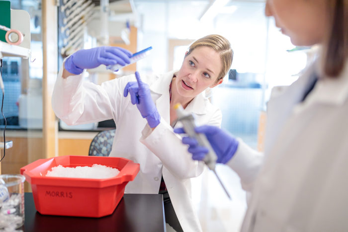
Samantha Morris, PhD, and her team at Washington University School of Medicine in St. Louis designed a cellular tracking system that can give scientists a new view of how cells develop. This "flight data recorder" for cells could one day help scientists guide cells along the right paths to regenerate certain tissues or organs, or help researchers understand the wrong turns some cells might take on their way to becoming cancerous.
Scientists at Washington University School of Medicine in St. Louis have developed a new tool described as a “flight data recorder” for developing cells, illuminating the paths cells take as they progress from one type to another.
Scientists hope to one day be able to take skin cells from a patient who needs a liver transplant, for example, and guide the skin cells along a known path that will result in a new liver. Without this cellular tracking device, researchers are able to study the original cells and the final cells in great detail — but the pathways cells take to reach their destinations have been largely unknown.
The study is published Dec. 5 in the journal Nature.
“There is a lot of interest in the potential of regenerative medicine — growing tissues and organs in labs — to test new drugs, for example, or for transplants one day,” said senior author Samantha A. Morris, PhD, an assistant professor of developmental biology. “But we need to understand how the reprogramming process works. We want to know if the process for converting skin cells to heart cells is the same as for liver cells or brain cells. What are the special conditions necessary to turn one cell type into any other cell type? We designed this tool to help answer these questions.”
According to the researchers, the tool could reveal cellular “reprogramming” routes that might involve reverting skin cells back to different types of stem cells that could then mature into a new liver or other vital organ. Among many potential uses, the tool also could be applied in cancer research, recording the wrong turns normal cells might take to develop into tumors.
Using their flight data recorder, the researchers performed experiments that uncovered some surprising details about the specific routes taken by cells that successfully completed their flight paths. The new information may help researchers identify the best “pre-flight” conditions to expose the cells to in order to increase the likelihood that their journeys will be successful. The scientists want to file the right flight plan, so to speak, so all of the cells arrive at the correct destinations.
“Right now, cell reprogramming is really inefficient,” Morris said. “When you take one cell population, such as skin cells, and turn it into a different cell population — say intestinal cells — only about 1 percent of cells successfully reprogram. And because it’s such a rare event, scientists have thought it is likely to be a random process — there is some correct set of steps that a few cells randomly hit upon. We found the exact opposite. Our technology lets us see that if a cell starts down the right path to reprogramming very early in the process, all of its related sibling cells and their descendants are on the same page, doing the same thing.”
The technique harnesses the natural properties of a virus that inserts tiny DNA “barcodes,” called “CellTags,” into each cell. As the cells divide, their unique barcodes are passed down to all their descendant cells. At several set time points during the 28-day cellular reprogramming window, new barcodes are added and a sample of the cells are analyzed to see what they’re doing at that waypoint. The CellTagging technique keeps track of which cells share common ancestors and how far back that common ancestor is found in the lineage, like a family tree. Indeed, beyond single flight tracking, the tool lets Morris and her team build complex family trees of cells, where successfully reprogrammed cells could be traced all the way back to their early ancestors.
Morris said their study suggests that the state the cell is in at the time it receives the instructions to reprogram has already set the stage for whether it will be successful or not. This contradicts assumptions that a cell goes in many different directions when it is first instructed to reprogram.
“If we can unlock that cocktail of initial conditions that primes cells to be successful in their reprogramming, we can transform cells into the type we want with much higher efficiency,” she said. “We’d like to reach 100 percent efficiency. That would be really exciting for the field of regenerative medicine.”
The researchers already may have identified one ingredient in the cocktail. They found that if a certain gene, called Mettl7a1, was turned on in cells, they were three times as likely to successfully reprogram compared with cells in which this gene is inactive. According to Morris, another interesting finding was that the cells that were not successful in their reprogramming didn’t just end up all over the map. They appeared to converge at the same dead end, tending to revert back to look like the original cell type.
“We see a single dead end right now, but as the technology improves, we think there’s a possibility we will see multiple outcomes,” she said.
The researchers studied mouse skin cells, exposing them to a molecular cocktail that triggers the DNA of the cells to open up, turn on new genes and reprogram skin cells into another cell type called induced endoderm progenitors.
Such cells give rise to both liver cells and to cells that make up the small intestine. In one example of a potential application, Morris wants to grow mini-guts to help the study of premature infants with a condition called short bowel syndrome. Babies born too early are at high risk of a disease that results in the death of tissue in the intestine. To stop the damage, some babies require surgery to remove that portion of the bowel, resulting in a shorter bowel.
“Since part of the intestine has been removed, these infants lose their ability to absorb nutrients,” Morris said. “If we eventually can grow replacement intestine from the human version of these cells, that would be an amazing advance.”
Morris worked with Washington University’s Office of Technology Management to patent the technology. Several research teams worldwide already have taken an interest in using the Morris lab’s CellTagging method in their own investigations.
“We are pleased and encouraged to see a number of cancer research groups adopting this technology in their labs,” Morris said. “With this method, they can find out which cells become the cancer and trace their ancestors back in time to see what they were doing early on.”


