Scientists map how human retinal cells relay information to brain
Study may advance understanding of eye diseases involving retina
 Getty Images
Researchers at Washington University School of Medicine in St. Louis report that specific types of retinal cells that carry the vast majority of visual signals from the human retina to the brain efficiently process and compress that information so it can be transferred. The scientists learned this by monitoring the activity of human retinal cells while retinas were being exposed to movies involving changing scenery.
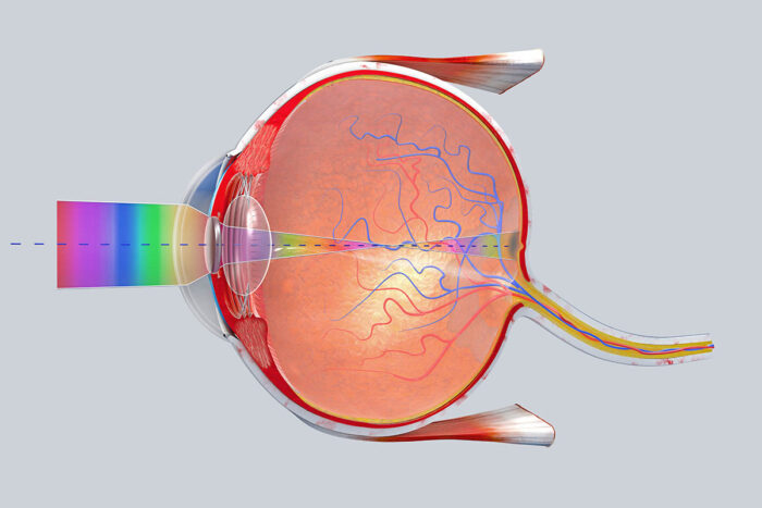
To understand how we see the world and how diseases such as age-related macular degeneration and glaucoma impair vision, scientists need to understand how the retina communicates vision signals to the brain.
Previously, researchers have worked primarily with retinal cells from animals. But a new study from scientists at Washington University School of Medicine in St. Louis details the researchers’ work with cells from human retinas and how those cells convert visual signals carried by the optic nerve to the brain, which then interprets the signals into sight.
In the study, published June 12 in the journal Neuron, the researchers report that specific retinal ganglion cells that carry the vast majority of visual signals from the human retina to the brain efficiently process and compress that information so it can be transferred. The scientists learned this by monitoring the activity of human retinal cells while retinas were being exposed to movies that caused those cells to transmit signals as the scenery changed.
“If we want to learn how to correct vision deficits in people who have retinal diseases, as well as problems like glaucoma that damage the optic nerve, we need to know how these ganglion cells convert and transmit signals,” said principal investigator Daniel Kerschensteiner, MD. “Visual systems are adapted to specific environments and behavioral demands, and these ganglion cells have adapted to those needs over millions of years of evolution. Although we share certain aspects of our visual systems with animals, if we want to really understand and restore vision in people, we need to better understand how human ganglion cells encode and relay visual information.”
Kerschensteiner, a professor of ophthalmology in the John F. Hardesty, MD, Department of Ophthalmology & Visual Sciences, and a professor of neuroscience and of biomedical engineering, led the study of retinal cells from human eyes that had to be removed due to ocular cancer. Although the scientists used retinas from only four human eyes, the researchers were able to draw conclusions by studying the activity of hundreds of ganglion cells in each eye. The axons of those cells join together to form the optic nerve.
The researchers focused on the two main cell types that transmit visual information, each of which comes in two varieties — one that responds when light increases and one that responds when light decreases.
Kerschensteiner’s team performed computer analysis of the cells’ activity and found that in every case, these cells transmitted the maximum amount of visual information using the minimum amount of energy. By using computer modeling to tweak how the cells behaved, the scientists were able to show that the ganglion cells were efficient. But whenever they made those adjustments on the computer, signals from the cells became less efficient.
“The optic nerve is a real bottleneck in the visual system,” he explained. “And it appears from our analysis that these cells evolved to compress visual information to get it through this narrow bottleneck. Patients with eye diseases, such as glaucoma, have difficulty doing that, and to correct those deficits, we need to better understand how healthy cells function. We’re just beginning to gain that understanding.”


