mRNA vaccines follow unconventional immune path to destroy tumors
Insight from study in mice could guide next-generation cancer vaccine development
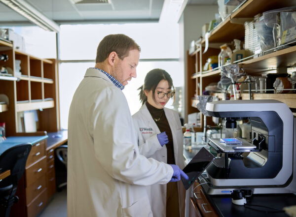
 Sara Moser/WashU Medicine
WashU Medicine researchers have described how mRNA cancer vaccines engage the immune system, through an unconventional pathway involving two subsets of immune cells called dendritic cells.
The advent of mRNA vaccines against SARS-CoV-2 in 2020 changed the course of the COVID-19 pandemic. Now, the Nobel-prize–winning technology is being adapted to fight cancer, with mRNA vaccines in clinical trials for melanoma, small cell lung cancer and bladder cancer, among others, opening the door to new ways of preventing and treating the disease.
Scientists assumed that one specific immune cell subtype was required for mRNA vaccination to activate the immune system. But researchers at Washington University School of Medicine in St. Louis show in a new study in mice that even without these cells, the mRNA vaccine still triggers strong cancer‑killing responses. That’s because, they found, a cousin to this subtype of immune cell can also stimulate anti-tumor immune activity — an unexpected finding given that this related subtype is not involved in responses to other vaccines.
The findings are published April 15 in Nature, offering a deeper understanding of how the immune system responds to mRNA vaccination and guiding the optimal design of a cancer vaccine.
“There is a lot of interest in applying the mRNA vaccine approaches used during the COVID-19 pandemic to the problem of inducing anti-tumor immunity,” said senior author Kenneth M. Murphy, MD, PhD, the Eugene Opie Centennial Professor of Pathology & Immunology at WashU Medicine. “By dissecting which immune cells are involved and how they coordinate the response, we’re offering vaccine developers some additional mechanistic insights to consider in their goal of optimizing these vaccines against tumor proteins.”
Murphy also is a research member at Siteman Cancer Center, based at Barnes-Jewish Hospital and WashU Medicine.
Unconventional immune pathway
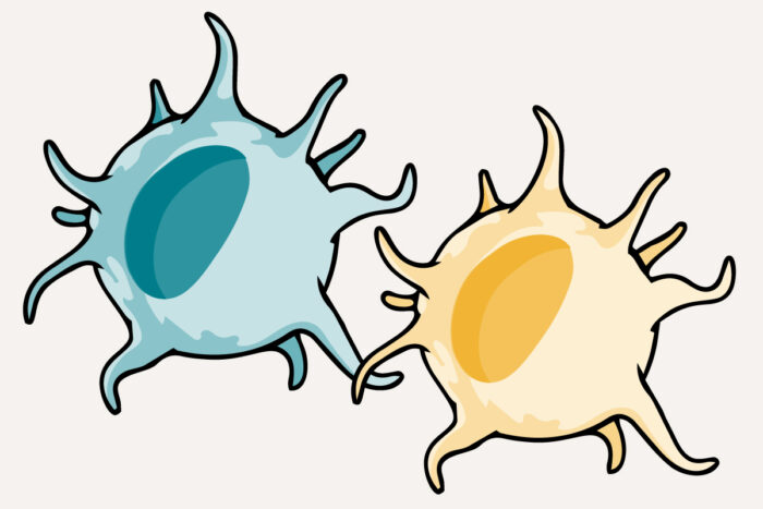
mRNA vaccines work by delivering instructions, in the form of messenger RNA biomolecules, for immune cells to produce bits of protein that trigger the immune system to destroy cells bearing these proteins. So-called dendritic cells produce the protein bits from the mRNA instructions, and T cells — another immune cell — are the ones that seek and destroy. mRNA vaccines can be designed to generate protein bits unique to a tumor so that T cells eliminate those cancerous cells.
cDC1, a classical type 1 dendritic cell, has long been known to be an effective teacher, priming T cells to attack cells infected by a virus. But less is known about how T cells become activated after an mRNA vaccine, whether against a virus or a tumor. In collaboration with the study’s co-corresponding author William E. Gillanders, MD, the Mary Culver Professor of Surgery at WashU Medicine, Murphy and members of his lab used mouse models that lacked cDC1 or a related cell subtype known as cDC2 to tease out the role that different groups of dendritic cells play in priming T cells after mRNA cancer vaccination.
Gillanders, a physician-scientist and surgical oncologist who also has developed an investigational vaccine against triple-negative breast cancer, treats patients at Siteman Cancer Center.
As part of the research, the scientists found that mice immunized with an mRNA vaccine generated strong T-cell responses even in the absence of cDC1s. In addition, they found that immunized mice without cDC1s were able to clear sarcoma tumors — cancers that develop in connective tissues such as fat, muscle, nerves, blood vessels, bone and cartilage. This indicated that some other cell type must be stimulating the T-cell response.
Indeed, their study found that cDC2s also participate in generating an immune response from T cells and preventing tumor growth. The study also found that T cells turned on by cDC1s and cDC2s each showed slightly different molecular “fingerprints.” These differences could help scientists design better versions of vaccines in the future.
Similarly, immunized mice lacking cDC2s and mice that had both cell subtypes produced an immune response and rejected tumor growth, demonstrating that mRNA vaccination uses both dendritic cell subtypes to stop cancer.
Further investigation of cDC2s suggested they activate T cells through an outsourcing process that relies on other cells to use the mRNA instructions to make the protein, chop it up and present small fragments on its surface. Once the protein is processed and presented, those cells then transfer the membrane complex that holds the fragment in place on the cell’s surface to the cDC2 to engage with the T cells — through an already-known process referred to as “cross dressing.”
“This work uncovers a new way mRNA vaccines engage the immune system — through both cDC1 and cDC2 — which helps explain their power and gives researchers concrete targets for making future mRNA cancer vaccines more effective,” said Gillanders. “It could improve vaccine formulation and dosing, potentially explain why some patients respond better to vaccines than others and guide strategies for making vaccines more effective.”