
Gut bacteria linked to malnutrition may pass to younger generations
Mouse study finds inflammatory microbes in mothers that affect fetal growth and the intestinal health of offspring
 Alexandra Byrne, PhD
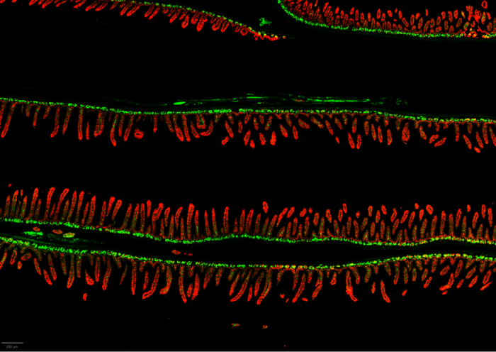
WashU Medicine researchers show how a disease of the small intestine related to malnutrition can be passed from mother to offspring. In a mouse study, they identify bacteria responsible for inflammatory signals that can damage the intestinal lining (labeled in red) and lead to increased cell division (labeled in green), a marker of injury to the tissue.
A new study led by researchers at Washington University School of Medicine in St. Louis suggests that an intestinal disorder linked to malnutrition and stunted growth may be transmitted from one generation to the next via the small intestinal microbiome. Analyzing mouse models of the disorder using bacteria cultured from children who themselves suffer from stunting and its detrimental effects, the researchers identified specific bacteria responsible for the inflammatory chemical signals that are characteristic of the disease, which damages the lining of the intestine and impairs nutrient absorption.
Published online in Nature Microbiology, the study provides evidence that mitigating inflammatory bacterial strains and blocking their transmission from mother to baby could serve as future treatment and prevention strategies for the disorder, known as environmental enteric dysfunction.
“The gut microbes in the small intestine make up a largely unexplored ecosystem — a ‘terra incognita’ — because they are difficult to sample. However, this study demonstrates how this ecosystem is important to the healthy growth and development of children,” said senior author Jeffrey I. Gordon, MD, the Dr. Robert J. Glaser Distinguished University Professor at WashU Medicine.
Importantly, the study showed how bacteria in a mouse mother’s small intestine affected intrauterine development of the offspring before birth, demonstrating that the effects of this disease extend beyond the walls of a mother’s gut to the developing fetus, according to Gordon, who also directs WashU Medicine’s Edison Family Center for Genome Sciences & Systems Biology.
“The study raises the question of whether manipulating the microbes in the small intestine either prior to or during pregnancy could help foster normal pre- and postnatal growth of their offspring,” Gordon said. “We are hopeful that this research could help us identify ways to break the vicious cycle of intergenerational malnutrition.”
How malnutrition may be passed to future generations
Many children around the world who suffer from malnutrition and stunted growth have an inflammatory condition of the small intestine called environmental enteric dysfunction, or EED. The disorder damages the lining of the small intestine and impairs absorption of nutrients from the diet. Such children also have poor immunity and deficits in cognitive development and growth (stunting) — problems that follow them throughout their lives even if their malnutrition is treated with standard therapeutic foods.
Working with collaborators at the International Centre for Diarrhoeal Disease Research in Bangladesh (icddr,b), Gordon and his colleagues analyzed samples from participants in the Bangladeshi Environmental Enteric Dysfunction Study, in which 525 malnourished children who averaged 18 months of age were treated with a standard therapeutic food that included milk, eggs, vitamins and minerals. Those children who did not improve despite the supplemental nutrition underwent an endoscopy procedure — with permission from their parents — to sample the tissue lining the gut and the bacteria present in their upper small intestine. The researchers’ earlier analysis of these samples identified 14 types of bacteria in the small intestine linked to stunted growth and inflammation.
In the new study, the researchers studied mice fed a diet representative of foods commonly eaten in the Mirpur district of Dhaka, Bangladesh, where the clinical trial participants lived. One group of these mice, born under sterile conditions without any gut bacteria, were colonized with carefully curated collections of small intestinal bacteria cultured from the children with malnutrion. This collection of human gut bacteria was shown to induce inflammation in the intestines of nonpregnant recipient female mice. A second group of mice, fed the same way, were given a different collection of gut bacteria from these children that didn’t induce inflammation, which served as a control. The researchers could not collect bacterial samples from healthy children’s small intestines as a control because it is unethical to perform endoscopies on healthy children.
Studying these mice, the researchers showed that the collection of inflammatory bacteria could be passed from the female mice to their offspring and that the harm begins in utero, before the bacteria are even present in the offspring. The mouse pups that received the inflammatory bacteria developed problems similar to those of the children with the disorder — stunted growth, markers of inflammation in their blood, and damage to their intestinal lining.
The researchers also found that when the separate groups of mice — those with inflammatory gut bacteria and those with non-inflammatory gut bacteria — were housed together, the inflammatory gut bacteria and their detrimental effects spread to the mice originally given the non-inflammatory bacterial collection.
The investigators found that the bacterial strain sampled from the children that contributed most to inflammation in the small intestines of the mice modeling their disease is called Campylobacter concisus. The researchers noted that a number of the inflammation-inducing bacterial strains identified in the small intestine are strains typically found in the mouth. In the mouth, they’re not known to cause problems but when then establish residency in the small intestine, they function as pathogens, suggesting that the surrounding environment, including neighboring bacteria, is key in determining whether a strain of bacteria is helpful, harmful or neutral in its effects.
In fact, when C. concisus was the only bacterial strain administered to germ-free mice — meaning mice with no gut bacteria of their own — the microbe didn’t cause disease or even survive very well, according to first author Kali M. Pruss, PhD, an instructor of pathology and immunology in Gordon’s lab at WashU Medicine.
“It highlights how nuanced these interactions are between different bacteria and how they contribute to disease in some contexts but not others,” Pruss said.
Gordon and his team are continuing their research to understand the complex and far-reaching effects of small intestinal gut bacteria. Their goal is to identify safe and effective ways to shift an inflammatory gut microbiome in mothers with EED towards one that not only reduces the damaging inflammation and malnutrition but also promotes establishment and maintenance of a healthy, stable gut microbial environment in their offspring.