New AI tools could help eye doctors diagnose retinal disease faster
Programs that analyze eye images could also speed up drug trials, study finds
 Getty Images/Zucks Liu
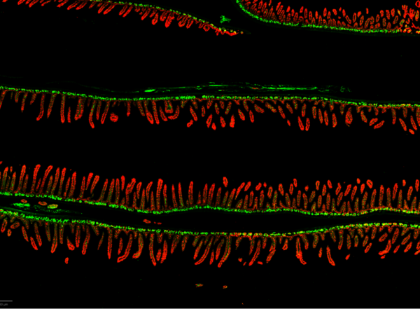
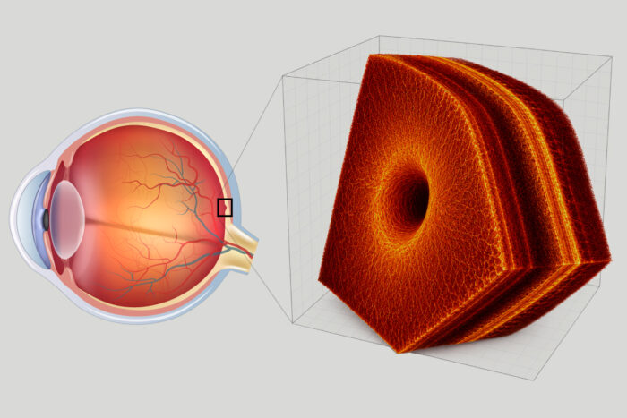
Researchers at WashU Medicine have developed an experimental AI system designed to interpret 3D images of the eye’s retina (thin yellow layer, left) for disease diagnosis. Each 3D image of the retina (inset at right) is composed of hundreds of 2D slices.
Non-invasive eye scans allow doctors a zoomed-in, three-dimensional look beneath the eye’s surface without causing discomfort or pain to the patient. Used routinely in clinics worldwide, the scans produce detailed views of individual layers of the eye’s interior to help diagnose conditions that threaten vision. But with that level of precision comes a flood of data — hundreds of images per scan that physicians have to review manually, a time-consuming process that is vulnerable to human error.
Now, researchers at Washington University School of Medicine in St. Louis, in collaboration with colleagues at the University of Washington in Seattle and Genentech, Inc., have developed an experimental artificial intelligence (AI) system that can speed the scan review process and help doctors spot subtle signs of eye disease sooner. The technology, called OCTCube-M, includes a family of three AI models that are designed to read and interpret 3D images of the eye’s retina as well as other types of eye scans.
In a new study, the researchers found that, compared with older models, the new AI system more accurately identified eight different retinal diseases, including age-related macular degeneration, a common disease that damages the retina and is the leading cause of blindness in people over 50. It also was more accurate in its predictions of how fast a severe form of this condition, called geographic atrophy, would progress.
The findings describing the technology in its research stage were published recently in Nature Biomedical Engineering.
“Today’s eye scans provide physicians an unprecedented, highly detailed view of the inside of the eye, revealing structures and subtle changes that would otherwise go undetected,” said the study’s co-corresponding author Aaron Lee, MD, the Arthur W. Stickle Distinguished Professor of Ophthalmology and Visual Sciences and head of the John F. Hardesty, MD Department of Ophthalmology & Visual Sciences at WashU Medicine. “But we still lack the tools to help physicians process the volume of generated images. Our AI system has the potential to empower physicians to make faster diagnoses, tailor treatment more precisely and design clinical trials that bring new therapies to patients faster.”
Additionally, the study showed that the model could infer health risks beyond the eye, predicting outcomes such as heart attack, stroke and kidney failure based solely on retinal imaging. The tiny blood vessels in the retina are anatomically and developmentally the same as those in the kidney, and the processes that lead to plaque buildup inside the walls of blood vessels that feed the heart and brain also leave signatures in the eye.
“The model has the potential to turn a simple eye exam into a powerful tool for helping to detect illness beyond the eye,” said Lee. “It opens the door to earlier detection, more precise monitoring and potentially better outcomes for patients who might otherwise go undiagnosed until their disease is far more advanced.”
A diagnosis needle in a haystack of data
At least 2.2 billion people worldwide have vision impairment, according to the World Health Organization. The imaging method known as optical coherence tomography has transformed the diagnosis and care of conditions that cause vision loss by generating, via a single and swift scan, hundreds of cross-sectional images that together form a detailed, 3D picture of the retina and the optic nerve. It can reveal early signs of different eye diseases such as glaucoma, macular degeneration and diabetic retinopathy, among other conditions.
AI, meanwhile, has become a powerful tool for processing large medical datasets, and Lee and colleagues have made notable contributions to the field. Several years ago, they published, in Nature, results describing a model that is better at diagnosing eye disease in two-dimensional retinal images compared to older models.
Because the model was trained on 2D tomography images, the researchers sought to determine if adding 3D tomography images could further improve disease diagnosis and prognosis. Because disease often extends in all three dimensions around the fovea, a small pit in the center of the retina responsible for the sharp, detailed vision required to read text and recognize faces, they hypothesized that training models on 3D images would provide more complete and accurate views of the tissue. To that end, more than 26,000 3D optical coherence tomography images comprising 1.62 million individual retinal slices — cross-sectional images of the retina — were used to train OCTCube-M.
When compared to the model trained on 2D images, OCTCube-M more accurately identified six of the eight retinal diseases by about four to six percentage points. That translates to the tool finding 43 to 60 additional cases out of every 1,000 individuals with eye disease. This was true across scans taken from individuals at multiple clinical sites, imaging modalities and diverse patient populations.
The eight diseases identified by the model include serious conditions that primarily affect the back of the eye, including the retina and optic nerve. Together they are the leading causes of vision loss and are linked to other conditions such as diabetes, hypertension and cardiovascular disease.
The researchers, including Cecilia S. Lee, MD, the Jane Hardesty Poole Distinguished Professor in ophthalmology and visual sciences at WashU Medicine; Sheng Wang, PhD, an assistant professor in the Paul G. Allen School of Computer Science & Engineering at the University of Washington; and Miao Zhang, PhD, a senior AI scientist at San Francisco-based biotech company Genentech, then adapted the 3D model by adding data from two other eye imaging techniques — infrared retinal imaging and fundus autofluorescence imaging.
By combining optical coherence tomography with one or both of the other imaging types, the AI models can construct a more complete view of the eye and a deeper understanding of what’s happening inside, Aaron Lee explained. Indeed, the model trained on all three imaging types excelled at predicting the growth rate of the severe form of macular degeneration, geographic atrophy, significantly outperforming the current state-of-the art model that trains only on fundus autofluorescence images of the retina by an average of nearly 50%.
Geographic atrophy affects about 5 million people worldwide, and there are few effective treatment options. By providing information on the growth rate of the condition, Lee and colleagues’ tool could effectively detect and classify the stage of the illness, information that researchers could use to design better clinical trials of potential therapies for the disease.
“By better predicting how fast disease will worsen, we can run smaller, more efficient studies,” Lee said. “That could lower costs, shorten the time it takes to test new therapies, reduce the number of people exposed to treatments that don’t work and help effective drugs reach patients sooner.”
Next, the WashU Medicine researchers will train OCTCube-M with larger datasets encompassing more patients, more diseases and even more types of imaging data to continue improving upon it.